Big Picture
The pterygopalatine fossa is the region between the pterygomaxillary fissure and the nasal cavity. The fossa accommodates branches of the maxillary nerve [cranial nerve (CN) V-2], the pterygopalatine ganglion, and the terminal branches of the maxillary artery.
The pterygopalatine fossa is an irregular space where neurovascular structures course through to the nasal cavity, palate, pharynx, orbit and face (Figure 22-1A and B). The neurovascular structures enter and exit the fossa through the following boundaries:
- Anterior boundary. Posterior surface of the maxilla.
- Posterior boundary. Pterygoid processes and the greater wing of the sphenoid bone, with openings for the following structures:
- Foramen rotundum for CN V-2.
- Pterygoid canal for the nerve of the pterygoid canal (Vidian nerve).
- Pharyngeal (palatovaginal) canal for the pharyngeal branch of CN V-2.
- Medial boundary. Perpendicular plate of the palatine bone containing the sphenopalatine foramen, which transmits the nasopalatine nerve (CN V-2 branch) and the sphenopalatine artery.
- Lateral boundary. Pterygomaxillary fissure, which communicates with the infratemporal fossa.
- Superior boundary. Greater wing and body of the sphenoid bone, with the infraorbital fissure transmitting the infraorbital nerve and the vessels in the orbit.
- Inferior boundary. Palatine process of the maxilla and the pterygoid process of the sphenoid bone with the greater and lesser palatine canals and foramina, which transmit the greater and lesser palatine nerves and vessels.
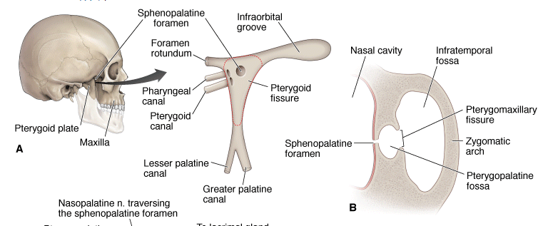
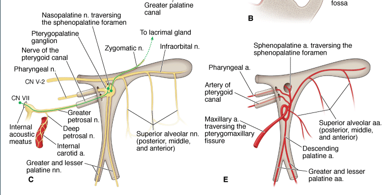
A. Outline of the pterygopalatine fossa. B. Axial section of the pterygopalatine fossa. C. and D. Nerves of the pterygopalatine fossa. E. and F. Arteries of the pterygopalatine fossa.
Innervation of the Pterygopalatine Fossa
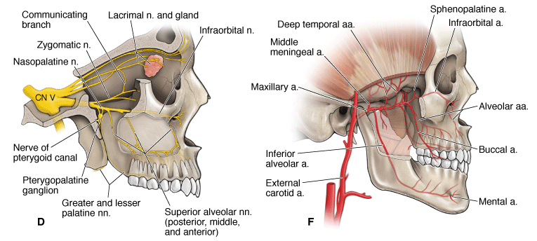
Branches of CN V-2 form most of the nerves that enter and exit the pterygopalatine fossa (Figure 22-1C and D). CN V-2 enters the fossa via the foramen rotundum and branches as follows:
- Posterior superior alveolar nerves. Enters the posterior superior alveolar canals, providing general sensation to the maxillary molar teeth and gingivae.
- Infraorbital nerve. Courses through the infraorbital fissure, groove, canal, and ultimately the foramen providing general sensation to the inferior eyelid, the lateral nose, and the superior lip. The infraorbital nerve also gives rise to the middle and anterior superior alveolar nerves, which supply the maxillary premolars, canines, and incisors, and the gingivae and mucosal lining of the maxillary sinus.
- Zygomatic nerve. Enters the orbit via the infraorbital fissure, dividing into the zygomaticotemporal and zygomaticofacial nerves, which supply the skin over the zygomatic arch and the temporal region. In addition, the zygomatic nerve communicates with the lacrimal nerve in the orbit and carries parasympathetic neurons from the pterygopalatine ganglion to the lacrimal gland.
- Pharyngeal nerve. Courses through the pharyngeal canal, supplying part of the nasopharynx.
- Greater and lesser palatine nerves. Descend through the palatal canals, emerging through the greater and lesser palatine foramina to innervate the hard and soft palates.
- Nasopalatine nerve. Traverses the sphenopalatine foramen, supplying the nasal septum before coursing through the incisive canal to supply part of the hard palate.
- Pterygopalatine ganglion. Lies inferior to CN V-2 and receives preganglionic parasympathetic neurons from the facial nerve (CN VII) via the greater petrosal nerve, traversing the pterygoid canal. The pterygopalatine ganglion sends postganglionic parasympathetic neurons to the lacrimal gland, via communicating branches between the zygomatic nerve and the lacrimal nerve (CN V-1), and the nasal and palatal glands, via the nasopalatine, greater palatine, and lesser palatine nerves.
- Sympathetics. Postganglionic sympathetic neurons from the superior cervical ganglion course along the internal carotid artery and give rise to the deep petrosal nerve. The deep petrosal nerve joins with the greater petrosal nerve (parasympathetics from CN VII) at the foramen lacerum to become the nerve of the pterygoid canal (Vidian nerve). The postganglionic sympathetic neurons course through but do not synapse in the pterygopalatine ganglion and inhibit lacrimal and nasal gland secretion.
The maxillary artery is a terminal branch of the external carotid artery, courses anteriorly through the infratemporal fossa, traverses the pterygomaxillary fissure, and enters the pterygopalatine fossa (Figure 22-1E and F). The maxillary artery supplies the maxilla, maxillary teeth, and palate before traversing the sphenopalatine foramen to terminate in the nasal cavity. The major branches of the maxillary artery in the pterygopalatine fossa are as follows:
- Posterior superior alveolar artery. Supplies the maxillary molar teeth.
- Descending palatine artery. Gives rise to the greater and lesser palatine arteries, which supply the soft and hard palates.
- Infraorbital artery. Supplies the maxillary tooth and skin of the face.
- Sphenopalatine artery. Traverses the sphenopalatine foramen to supply the nasal cavity.
Content 2
Content 3